The DU Lounge
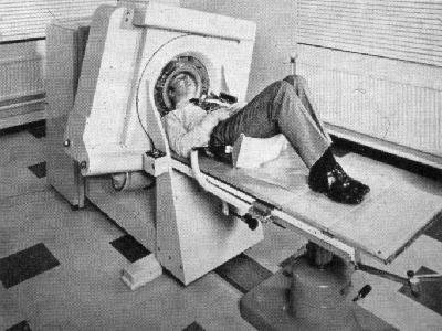
Related: Culture Forums, Support ForumsIf you ever wondered - "What does a CT scanner really look like without the shell"
 = new reply since forum marked as read
Highlight:
NoneDon't highlight anything
5 newestHighlight 5 most recent replies
= new reply since forum marked as read
Highlight:
NoneDon't highlight anything
5 newestHighlight 5 most recent replies

KT2000
(22,151 posts)also - the patient has no idea all of this is taking place during the scan.
mitch96
(15,804 posts)what was flying around their body.
Thanks for the neat pic...
m

Control-Z
(15,686 posts)Brother Buzz
(39,900 posts)I've had MANY scans: I had no clue there were spinning parts inside

Lars39
(26,540 posts)
LAS14
(15,506 posts)Lars39
(26,540 posts)
Marie Marie
(11,310 posts)mitch96
(15,804 posts)of the magnetic..Another neat factoid is that the magnet and coils are held in a big thermos bottle held at -454ºF/-270ºC...It uses liquid helium... You haven't lived till you have seen an MRI unit quench the magnet and blow off all the helium..... So much "smoke" it looks like it's on fire! The coils produce a bucket load of heat and have to be kept cool. Same with the CT scanner but not as hot. If I remember my XRay physics an xray tube produces 1% Xray and 99%heat.. not too efficient..
ok end of class...
m
KT2000
(22,151 posts)I had one done years ago. They injected the dye and I had to wander around until it hit my brain. When I went into the room for the scan I was hit with how clean the air was (I am sensitive to such things) and mentioned it to the tech. I noticed two large and loud air filters on the ceiling. The tech said that was for the purpose of keeping the room cool enough to run the scan. Does that sound right to you?
mitch96
(15,804 posts)gigercounter but much more complicated.. it's computerized so it's GOT to be complicated. PET scans (positron emission tomography) show function of organs and tumors. If they pick up the radioactive isotope (the stuff they injected) and it "functions" normal your good If the function is abnormal there could be a problem...Xray and CT mostly show structure.... like seeing a broken leg.... One piece is normal two pieces it's broken.. MRI can show structure AND function also...
Way cool stuff and doing the tests and working on the million dollar machines was lots of fun.
and And AND they paid me to play with the big toys.. I loved doing CT/MRI... You could really geek out on them things. We did a lot of good for people and showed problems way before they showed up on other tests... Just the problem of using all them pesky xrays though... MD's did not give a shit about patient exposure.... Lots of the MD's were into "rule out" and CYA diagnosis..Lucky the mfg saw that problem and started using technology to reduce patient exposure to a point where it was down to safe levels relatively speaking..
No radiation is good radiation.... and I'll leave it at that..
m
KT2000
(22,151 posts)Maybe you could explain something else. I have had two MRI's one on the neck and one for lower back. When I had the neck MRI, there was a point where whatever energy it is, hit the problematic nerve. It felt like a super hot fire cracker fuse that went from my neck down the arm and hand to the thumb. That is where the problem was to begin with and after the MRI, the pain was gone.
The lower back also had the hot nerve episode without traveling anywhere else.
No doctor will explain what that was or even if that was supposed to happen. At this point if any doctor suggests an MRI on my brain, I will kindly refuse.
Glad they are reducing radiation. Years ago a friend did hospital inspections for nuclear departments. He said they all lied except the Catholic hospital that told the truth. They got written up and the liars got good reports!
mitch96
(15,804 posts)To my untrained mind it sounds positional. Just lying still for so long maybe kinda sorta pooched out the disk in your cervical spine. From what you wrote with the shooting hot pain down your arm sounds like a herniated disk putting pressure on the nerve going to your arm and hand and radial nerve. Nerve pain is hot...
The older MRI scanners would produce heat in some patients but not all would feel this. I had a crap load of MRI's and never got hot... Maybe I was not "wet" enough? Hope this helps.
The magnet aligns the water molecules in the body and the protons in the hydrogen atoms move when you hear the "bang"of the gradient coils. Each bang produces a bunch of dots. Enough dots any you have a picture. MRI physics is really voodoo stuff.So you do this a few thousand times and the the little buggers heat up. I don't have any problems having MRI's but I'd watch out for the contrast material they use. It's made from a toxic metal in very small doses. I always refused the contrast.
MRI's don't use ionizing radiation like xray... One thing that we noticed on the older machines was that we were in the magnetic field while at the control panel. You put a metal pen down and it would snap to align with the magnet, like a compass to the north pole. We found that if we worked daily on the machine for a month or so we would get short term memory loss. This was with young healthy tech's. We would have to write everything down or it was lost to the ethers. Go on vacation and in a day or so everything was fine... The MRI mfg's increased the distance from the magnet in the newer machines and the problem went away....
About checking the output of xray machines.... LOTS of lying goes on in hospitals.. Follow the money...
m
KT2000
(22,151 posts)You are the first person to answer a question I have asked doctors and others. I always have a feeling when I ask questions anymore, people don't answer because they are afraid of a lawsuit or something when I just wanted to understand something. As you say - money.
Again thanks!
mitch96
(15,804 posts)sometimes and many times they can't. So the easiest thing to do is not answer or answer a different question with a lot of medical terminology. Sounds impressive but not productive for the patient. I can't tell you how many times I've had a radiologist explain to a patient something and the patient has that quizzical WTF look on their face. I would try to bring it down to "normal speak" and relieve their apprehension. Lawyers do the same thing... They speak legalese and the customer does not know what the heck he/she said... And don't get me going on IRS speak.. totally baffling. I guess all professions want to make them selfs special with their own jargon
For me? you can say fecal encephaly or shit for brains. Most people understand the second. You can say pneumonoultramicroscopicsilicovolcanoconiosis or little specks of silicone in the lungs.
Take your pick...
m
I'll go with shot for brains, errr I mean trump!

csziggy
(34,189 posts)When my sister got her PhD, her doctoral thesis was on "Computed Tomography of the Brain." It was later published as a reference book for neurosurgeons - she compared old style slices of diseased brains to images made with the then new (about 1987) technology.
A few years later she was diagnosed with glioblastoma. She got lots of attention and some experimental treatment since so many neurosurgeons had used her book as a reference. Originally her prognosis was that she would live only three months after diagnosis - her friends gave her about twenty months of quality life.
One of life's ultimate ironies, there.
mitch96
(15,804 posts)translate rotate machine at NYU med ctr in New York City.. It was the 4th one in the US and the 6th one in the world. New stuff!!
I had broken my ankle in a motorcycle accident and was in a half cast. My boss wanted to give me something easy to do so they stuck me on the CT scanner... No body wanted to work on the machine.... I never looked back..
You were talking about slices of the brain, we took a frozen cadaver brain and had the brain cut into 10 mm sectons. Then we xrayed the sections and put the pic's side by side with the CT slice of the brain for a book that was being published Maybe your sister???
That old EMI machine did only brain scans. It was all mechanical. each slice has to mechanically moved to the next position with a little wheel and ruler and the scan restarted. Twenty min for each slice. It took almost 45 min to do a scan and the patient had to hold perfectly still. Then it took about 20-30 min to process the images... My fricken iPhone is more powerful than the racks and racks of computer hardware that scanner had. And yes, we scanned blind. We did not see the pic's for ½ hr after the patient left..
Machines got faster and faster. It was great being on the bleeding, leading edge of technology that everybody now takes for granted. God I have a ton of stories about those times.. Long time ago in a far away place.
Here is a pic of the old machine. The Medical museum at the Mayo Clinic has an old Emi scanner on display.. at least they had one years ago..
That whole thing around the patients head rotated. You can see the xray tube peaking out on the bottom. The tube would go back and forth perpendicular to the patients head, then the gantry would rotate Do this 8 or 10 times, go out and change the location with the little wheel and repeat. The "table control" is near the patients right elbow and thats how you moved the patient in and out The machine had to reference water before going thru tissue so the patients head is in a plexiglass box full of water with a rubber cap that went over the patients head.... It looked like an elephant condom. Yes they would break and douse the patient with water!!! Great fun for a 20 something tech eager to learn everything at once simultaneously together..
Like I said I'm an antique...m
csziggy
(34,189 posts)My sister got her PhD at University of South Florida in Tampa. Her soon to be husband helped with the photography but I don't know where most of the pictures were taken.
I've had MRIs and CT scans - I much prefer the CT ones. One of those saved my life.
After years of complaining about shortness of breath I was finally diagnosed with a defective aortic valve. At the time I was considered a low risk patient and the non-invasive transcatheter aortic valve replacement (TAVR) was only approved for high and medium risk patients. The cardiothoracic surgeon who did the tests to measure how inefficient my valve had gotten (40%) was part of a clinical trial to get TAVR approved for low risk patient and recruited me to join the study.
That gave me a 50% chance of not having to have my chest cut open so I went through all the approval process. The very last step was to get a CT scan to make sure my arteries were open enough to do the procedure. That was on a Thursday. The next Monday he called and told me "We have a problem. There is a mass on one of your kidneys." He hoped the kidney could be resected and I could stay in the study.
Off to a urologist who said no resection because of the location of the mass so they had to take the kidney out. But he did not want to do that with the bad valve. So I got kicked up to a higher risk level, got my new valve and a month later lost a kidney. The mass was cancerous and of the type that is usually not found until it spreads.
A friend told me that is a hell of a way to get the procedure I wanted!
mitch96
(15,804 posts)Very rare to spread and long term outlook is very good....
m
csziggy
(34,189 posts)I am over due to get scanned this year. It's been three years since they took it out and the doctor wanted me to get scanned every year for a minimum of three years. What with Covid and other things, I have not wanted to deal with it and put it off from this summer. Now that my life has stabilized, of course Covid has flared back up so I am putting it off some more. I may just wait until next spring when the pandemic has slowed down and it will be just before my next checkup with the urologist is schedules.
mitch96
(15,804 posts)You hear stories about radiation oncologists getting cancer, vascular surgeons getting blood vessel disease and nephrologist getting kidney stones. The disturbing thing is your sister knew exactly what was going on and what her prognosis was... You can take solace in the fact that her work helped others to see further by standing on the shoulders of giants. I salute her....
m
csziggy
(34,189 posts)But did not want to admit it until the tumor got so big it sent her to the emergency room. Various neurologists assisted over the year and a half before she went. They really wanted to help her survive. They learned a lot about treatment with the methods available in the early 1990s. I think much of what they learned helped them treat other patients.
Now there are other methods than surgery which are being explored and it is exciting to hear about those.
She was a brilliant researcher and I miss her still - she died in 1992.
LAS14
(15,506 posts)
localroger
(3,782 posts)CT scan is basically a very fancy X-ray. MRI uses the magnet and radio fields to make the atoms wobble, and detects the wobbles with radio receivers to determine what elements are present.
mitch96
(15,804 posts)
The Velveteen Ocelot
(130,537 posts)Sometimes you don't want to see the innards of things.
Marie Marie
(11,310 posts)mitch96
(15,804 posts)and we pulled the patient out quickly and then all the oil started dripping out.. what a mess...
Then you have to explain to a waiting room full of out patients that we are going to have to reschedule their test due to an "unforeseen" problem with the scanner...
The spinning thing don't spin no moe..
m